
01
Pattern
What type of change is happening?
Shedding, gradual thinning, recession, patchy loss, breakage, or another pattern.
A dermatologist-led evaluation designed to understand why your hair is shedding, thinning, or changing before a treatment plan is selected.
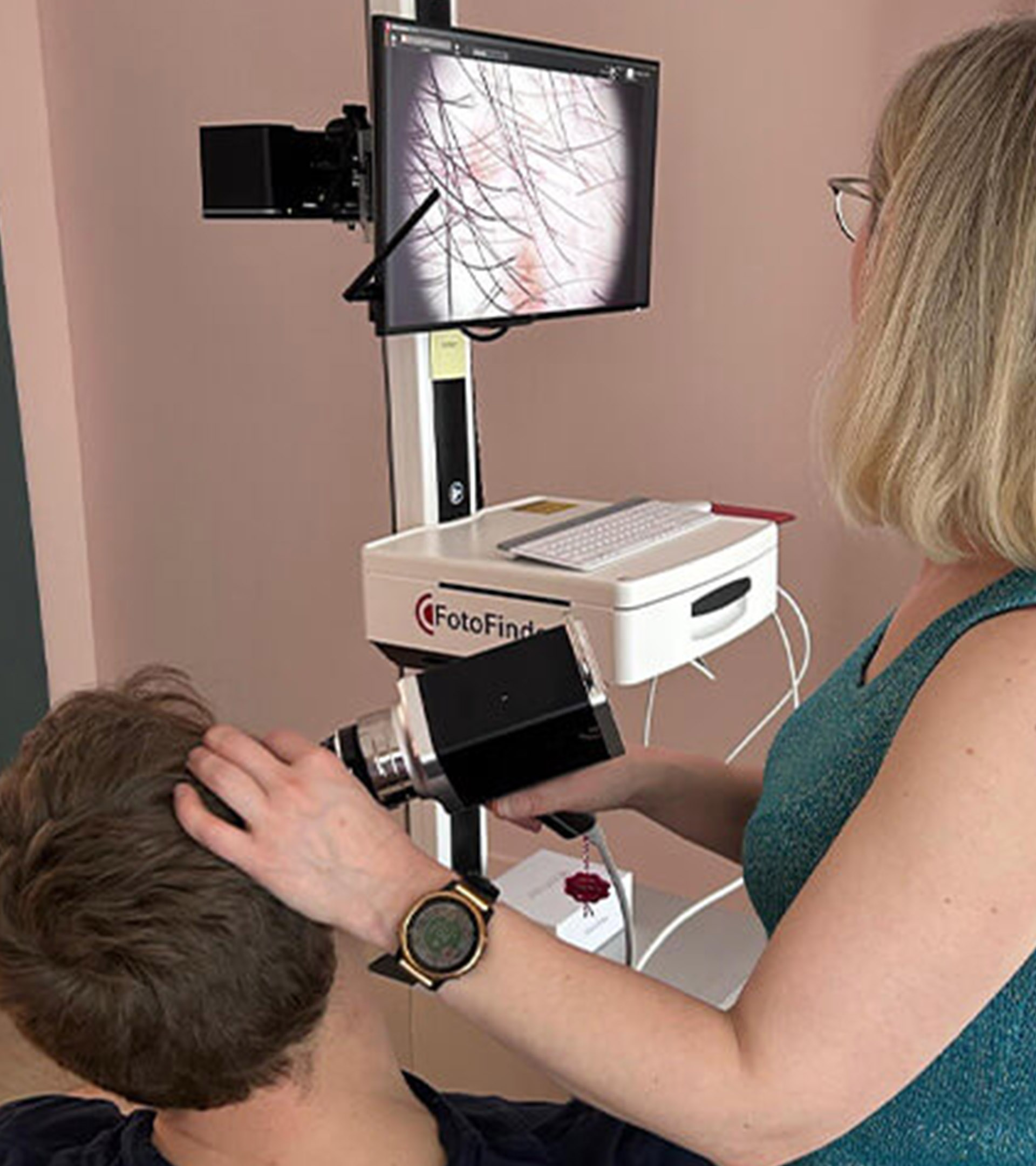
Your visit brings together medical history, hair and scalp examination, digital trichoscopy, and additional testing when it may provide clinically useful information.
Medical evaluation first. Treatment options are considered after the clinical picture is understood.
Hair shedding, thinning, and changes in density can look similar while reflecting very different processes.
The first goal is to identify which findings matter before deciding what — if anything — should be treated.
Similar symptoms do not always mean the same diagnosis.
Shedding, gradual thinning, recession, patchy loss, breakage, or another pattern.
Miniaturization, inflammation, scaling, altered follicular openings, or other findings.
Health, medications, nutrition, hormonal changes, weight changes, or hair practices may add context.
Activity and progression can influence treatment priorities and follow-up.
Once these questions are clearer, the consultation can move from concern to a structured clinical assessment.
See What Happens During the ConsultationHair loss evaluation is a structured medical process, not a single test.
History, examination, trichoscopy, and selective testing are brought together to understand what may be affecting the hair and scalp.
When did the change begin, how has it progressed, and is the main concern shedding, thinning, recession, breakage, or patchy loss?
Density, distribution, breakage, scaling, inflammation, and other visible findings help guide the next part of the evaluation.
Magnified imaging can reveal miniaturization, follicular openings, hair-shaft variation, and signs of inflammation.
Blood work or scalp biopsy may be considered when the findings raise a specific clinical question that needs clarification.
The information is interpreted as a whole to determine the most likely process and the appropriate next step.
Not every patient needs every test. The evaluation is guided by what the history, examination, and trichoscopy actually show.
Trichoscopy provides a magnified view of the hair, follicles, and scalp and can reveal findings that may not be obvious during visual examination alone.
These images are interpreted together with the patient's history and clinical examination. A trichoscopic image provides evidence — it is not a diagnosis by itself.
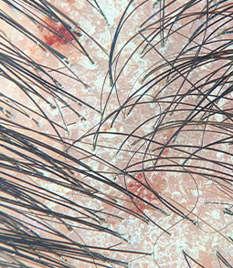
Psoriasis of the scalp. Scaling and focal bleeding seen on dermoscopy.
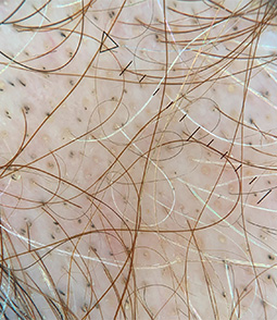
Black dots and yellow dots, as well as miniaturized hair – signs of active alopecia areata.

Miniaturized hair and thin pigtail re-growing hair in alopecia areata.
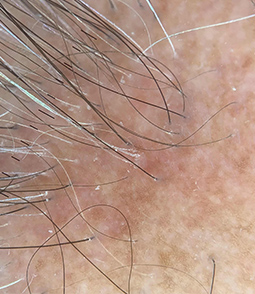
Lichen planopilaris / Frontal fibrosing alopecia. Dermoscopy demonstrates inflammation around hair follicles.
The appearance of a single image is not enough to establish a diagnosis. Similar findings may occur in different conditions and must be interpreted in clinical context.
Trichoscopy helps organize subtle findings into patterns that can support the clinical diagnosis.
Digital imaging can document how density and follicular distribution differ between areas of the scalp.
Variation in shaft thickness and the presence of progressively finer hairs can provide useful diagnostic information.
The appearance and preservation of follicular openings can help distinguish different hair-loss processes.
Redness, scaling, and perifollicular changes may provide clues when inflammatory or scarring disorders are being considered.
Stored digital images can create a reference point for comparing findings at future evaluations.
Not every patient with hair loss needs the same laboratory tests — and many do not need a scalp biopsy.
Additional testing is considered when it can help answer a specific clinical question or clarify the next step.
The history and examination help determine whether laboratory evaluation for a possible systemic contributor is appropriate.
When deficiency or anemia is clinically relevant.
When the history or symptoms suggest it may matter.
After relevant dietary or significant weight changes.
When supported by the clinical picture.
There is no single universal blood panel for every patient with hair loss.
Biopsy may be considered when the diagnosis remains uncertain or when clinical findings raise concern for an inflammatory or scarring process.
Microscopy can provide another layer of diagnostic information.
Especially when examination or trichoscopy shows inflammatory changes.
Histopathology may help characterize a process affecting the follicles.
The purpose of testing is not to collect more data. It is to obtain information that may change the clinical assessment or next decision.
Why Your History MattersHair changes are interpreted in the context of your health, timeline, medications, family history, nutrition, and hair practices.
These details can provide important clues — but they only become meaningful when they fit what is seen during examination and trichoscopy.
A possible trigger is not automatically the diagnosis. Context helps determine which factors deserve clinical significance.
Family history and age of onset can provide context when evaluating gradual thinning, density changes, or a changing hairline.
Illness, medication changes, hormonal changes, or other health events may be relevant when their timing corresponds with changes in the hair.
Restrictive diets, significant weight changes, or nutritional concerns may help determine whether additional evaluation is appropriate.
Traction, extensions, heat, and chemical processing can contribute to breakage or influence the pattern seen during examination.
History provides context. Examination provides evidence. The two are interpreted together before a clinical plan is developed.
What You Leave the Consultation WithThe purpose of the visit is not simply to collect information — it is to decide what the findings mean and what should happen next.
Depending on the clinical picture, the next step may involve treatment, additional evaluation, observation, or structured follow-up.
History, examination, trichoscopy, and available test results are interpreted together to identify the process that best explains the findings.
If another test could meaningfully clarify the assessment, the reason for that next step can be discussed.
When the clinical picture is sufficiently clear, treatment priorities and appropriate options can be discussed.
Follow-up may help determine whether the condition is stable, progressing, or responding to treatment over time.
The plan should reflect the diagnosis, the activity of the condition, the clinical findings, and the individual patient.
Once the evaluation identifies the likely cause, the next step is a diagnosis-specific plan for treatment and follow-up. Explore our broader hair loss care in NYC for the conditions we treat, medical therapies and additional treatment options.
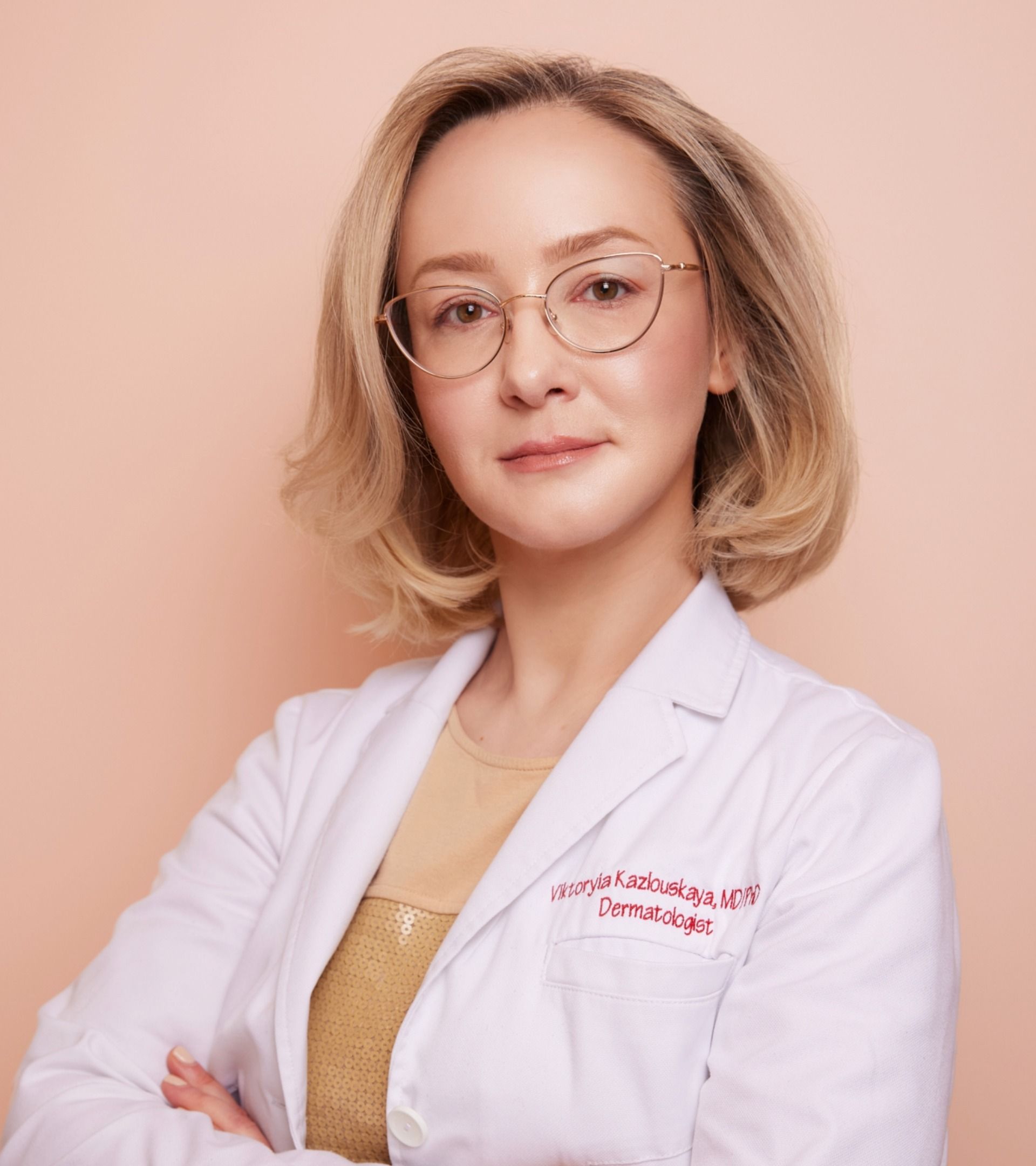
Board-Certified Dermatologist · Dermatopathologist
Hair loss evaluation requires more than identifying whether hair appears thinner.
The goal is to understand the pattern, examine the scalp and follicles, determine whether inflammation or another process may be present, and decide which additional information is actually useful.
“With hair loss, my first goal is to understand which process is actually affecting the follicles.”
Hair density, scalp findings, distribution, and pattern are evaluated in context.
Magnified follicular findings can add another layer of information to the examination.
When biopsy is needed, dermatopathology adds perspective to complex inflammatory or scarring hair-loss cases.
Physician-led medical hair loss evaluation
Additional perspective when microscopic evaluation matters
Clinical examination, trichoscopy and longitudinal assessment
You do not need to arrive with a diagnosis or a complete workup already performed.
The consultation is designed to determine which information is useful, what may need further evaluation, and what the appropriate next step should be.
No extensive preparation is required. It can be helpful to think about when the hair changes began, whether the problem is mainly shedding, thinning, recession, breakage, or patchy loss, and whether anything changed around the same time.
In most cases, the scalp should simply be in its usual condition. Avoiding heavy styling products, fibers, powders, or products that obscure the scalp can make examination and trichoscopy easier.
Yes, when available. Previous laboratory results, pathology reports, photographs, medication lists, and details of treatments already tried may help provide additional clinical context and avoid unnecessary repetition of testing.
No. Laboratory testing is selected according to the history, examination, shedding pattern, medical background, and suspected diagnosis. There is no single universal blood panel required for every patient with hair loss.
No. A scalp biopsy may be considered when the diagnosis remains uncertain or when examination and trichoscopy raise concern for an inflammatory or scarring process. Many hair-loss conditions can be evaluated without biopsy.
Digital trichoscopy is magnified imaging of the hair, follicles, and scalp. It can help document features such as hair shaft variation, miniaturization, follicular openings, scale, and signs of inflammation. The findings are interpreted together with the clinical examination.
Yes. Different processes can overlap. For example, increased shedding may occur in someone who also has pattern hair loss, and inflammatory disease may coexist with another form of alopecia. Recognizing overlap can change treatment priorities.
When the clinical picture is sufficiently clear, appropriate treatment options and follow-up can be discussed. If additional testing is needed first, the consultation can instead define what information should be obtained before finalizing the treatment plan.
You do not need to know why your hair is shedding, thinning, or changing before making an appointment.
The purpose of the consultation is to determine which findings matter, whether further evaluation is needed, and what the appropriate next step should be.
Physician-led hair and scalp evaluation at Dermatology Circle in New York City.