
Inflammatory Scarring Hair Loss · NYC
Lichen Planopilaris & Frontal Fibrosing Alopecia Treatment in NYC
LPP and FFA can permanently damage hair follicles. The first question is not simply how much hair has been lost, but whether inflammation is still active today.
These related immune-mediated forms of cicatricial alopecia may require careful pattern recognition, trichoscopy and, in selected cases, scalp biopsy.
The Clinical Priority
Recognize the pattern.
Determine activity.
Preserve follicles that remain viable.

01
Clinical Pattern
02
Disease Activity
03
Trichoscopy + Biopsy
04
Long-Term Stabilization
Why Timing Matters
Inflammation Can Lead to Permanent Follicular Loss.
Lichen planopilaris and frontal fibrosing alopecia belong to the lymphocytic spectrum of primary scarring alopecia.
Inflammation develops around the follicular unit and can progressively damage structures required for continued hair growth.
Once a follicle has been completely destroyed and replaced by scar tissue, treatment cannot simply recreate it.
A Different Measure of Success
Controlling inflammation
and preserving remaining follicles
can be meaningful treatment outcomes.
LPP vs FFA
The Distribution Often Gives the First Clue.
These conditions overlap biologically, but their visible patterns can be quite different.
Recognizing the distribution helps guide scalp examination, trichoscopy and decisions about further testing.
01
LPP
Lichen Planopilaris
LPP often presents with irregular or multifocal areas of scarring hair loss. Inflammation may be most visible around follicles at the active margin.
Pattern
Multifocal or irregular patches
Scalp
Perifollicular scale and erythema
Symptoms
Itching, burning or tenderness may occur
Other Clues
Signs of lichen planus may occur elsewhere
in some patients
02
FFA
Frontal Fibrosing Alopecia
FFA characteristically causes recession of the frontal and temporal hairline and frequently affects the eyebrows.
Pattern
Frontal and temporal recession
Eyebrows
Frequently involved
Trichoscopy
Loss of follicular openings,
inflammation and lonely hairs
Other Clues
Facial papules or body-hair loss
may occur in some patients
Clinical Context
FFA is closely related to LPP,
but its characteristic hairline pattern
gives it distinct diagnostic
and treatment considerations.
Disease Activity
Is Inflammation Still Damaging Follicles?
The amount of visible hair loss does not necessarily tell us how active the disease is today.
Active inflammatory areas and established scar may exist side by side.
Follicles Remain, but Inflammation Is Present.
The priority is identifying areas where treatment may still help protect follicular structures from further injury.
Perifollicular erythema
Perifollicular scale
Progressive loss or recession
Symptoms may be present
Active trichoscopic findings
Follicular Structure Has Already Been Lost.
Fully scarred areas have limited potential for regrowth, even when surrounding inflammation is successfully controlled.
Absent follicular openings
Smooth or shiny scalp
White or atrophic areas
Established hairline loss
Limited regrowth potential
Important
A relatively quiet scalp does not always mean inactive disease.
Symptoms alone cannot establish
whether inflammatory scarring alopecia has stabilized.
Diagnosis
Diagnosis Comes Before the Treatment Plan.
LPP and FFA may overlap with other inflammatory and nonscarring forms of hair loss.
Evaluation considers distribution, follicular openings, perifollicular inflammation, eyebrow involvement and whether another hair-loss process may be present at the same time.
Clinical Pattern
Multifocal loss or frontal recession?
Scalp Examination
Scale, redness, symptoms and established scar
Trichoscopy
Follicular openings,
erythema, scale and hairline clues
Disease Activity
Which follicles still show active inflammation?
Additional Testing
Biopsy or laboratory monitoring
when clinically appropriate
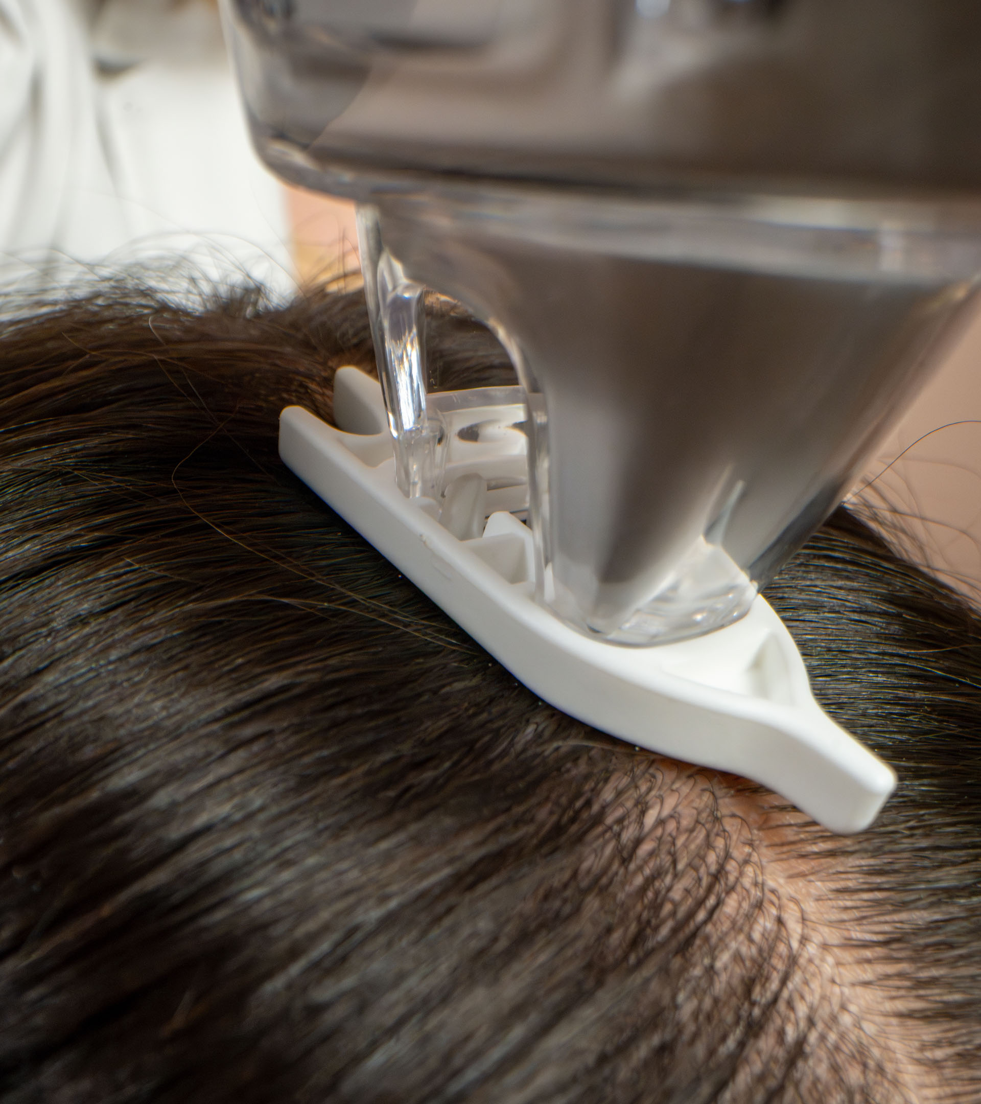
Scalp Biopsy + Dermatopathology
The Active Edge May Tell Us More Than the Scar.
When scalp biopsy is needed, where the sample is taken can matter.
A completely scarred area may mainly demonstrate the final structural damage. An active or transitional margin may reveal more about the inflammatory process that is still occurring.
Clinical examination and trichoscopy can help identify an informative site where follicles remain and inflammatory changes are still present.
Learn More About Scalp Biopsy
Clinical Pattern
Identify Where Disease Appears Active
Determine where follicular inflammation meets established hair loss.
Trichoscopy
Refine the Biopsy Site
Perifollicular scale, erythema and remaining follicular openings may help identify a useful sampling area.
Dermatopathology
Examine the Follicular Injury
Microscopy may demonstrate lymphocytic inflammation, fibrosis and follicular destruction.
Clinical Correlation
Match the Microscopy to the Patient
Histopathology is interpreted together with the clinical pattern, trichoscopy and disease course.
Dermatology at the Scalp.
Dermatopathology Under the Microscope.
Treatment Strategy
Control Active Disease Before More Follicles Scar.
There is no single treatment protocol for every patient.
Management depends on disease activity, extent, clinical pattern, medical context and coexisting hair loss.
Local Disease Control
Suppress Active Inflammation
Topical or intralesional corticosteroids and selected other local anti-inflammatory therapies may be used according to location and disease activity.
Progressive Disease
Escalate When Local Therapy Is Not Enough
Hydroxychloroquine, doxycycline and other systemic anti-inflammatory or immunomodulatory strategies may be considered when clinically appropriate.
FFA Considerations
Address the Hairline Pattern
In selected patients with FFA, 5-alpha-reductase inhibitors may form part of an individualized plan with appropriate counseling.
Overlapping Hair Loss
Treat Pattern Hair Loss Separately
Cicatricial alopecia can coexist with androgenetic alopecia. Minoxidil or other appropriate treatment may be added for the nonscarring component.
Reassessment
Document Change Over Time
Symptoms, clinical examination, standardized photography and trichoscopy may help determine whether disease is stabilizing.
What About Hair Transplantation?
Transplantation may be discussed
in carefully selected patients
after disease has become clinically stable.
Because cicatricial alopecia can reactivate and graft survival may be reduced, candidacy requires cautious individualized assessment.
Long-Term Management
Stabilization Is a Clinical Outcome.
With scarring hair loss, meaningful improvement is not always dramatic visible regrowth.
Baseline
Document Pattern & Activity
Control
Treat Active Inflammation
Reassess
Compare Symptoms, Examination & Trichoscopy
Maintain
Watch for Stability or Reactivation

Dermatology + Dermatopathology
Scarring Hair Loss Requires Diagnostic Precision.
The challenge is not simply recognizing that hair has already been lost. It is determining whether inflammation remains active and which follicles may still be preserved.
Dr. Viktoryia Kazlouskaya is board-certified in dermatology and dermatopathology. This dual perspective is particularly relevant when clinical findings, trichoscopy and scalp biopsy need to be interpreted together.
The goal is a treatment strategy built around diagnosis and disease activity, rather than a standardized menu of hair-loss procedures.
Practice Principle
The more permanent the potential damage,
the more important the diagnosis becomes.
Hair Loss Care · Manhattan
LPP & FFA Care on the Upper East Side.
Physician-led evaluation for inflammatory and scarring hair loss in a private Manhattan practice.
LPP & FFA FAQ
Questions Patients Often Ask.
The central questions are whether inflammatory disease remains active and which follicles remain viable.
01 What is lichen planopilaris?
Lichen planopilaris is an inflammatory form of primary scarring alopecia that affects hair follicles, most commonly on the scalp. It can lead to permanent follicular loss.
02 What is frontal fibrosing alopecia?
Frontal fibrosing alopecia is a related lymphocytic scarring alopecia characterized by recession of the frontal and temporal hairline. Eyebrow loss is also common.
03 Is FFA the same as LPP?
FFA is closely related to LPP and is commonly considered part of the same disease spectrum, but its clinical distribution and some treatment considerations differ.
04 Can hair grow back after LPP or FFA?
Regrowth depends on whether the follicular structure remains viable. Follicles that have been completely destroyed and replaced by scar tissue cannot reliably produce new hair.
05 How can you tell if the disease is active?
Activity may be suggested by progression, perifollicular redness or scale, symptoms and trichoscopic findings. No single symptom alone reliably establishes activity.
06 Can LPP or FFA stop progressing?
These conditions can become clinically stable, either with treatment or during their natural course, but activity can also persist or recur. Long-term follow-up may therefore remain important.
07 Do I need a scalp biopsy?
Not every patient requires biopsy. It may be especially useful when the diagnosis is uncertain or when histopathology may influence treatment. When performed, selecting an informative biopsy site matters.
08 How are LPP and FFA treated?
Treatment is individualized. Local corticosteroids, intralesional corticosteroids and systemic anti-inflammatory or immunomodulatory medications may be considered depending on disease activity, extent and medical context.
09 Is minoxidil a treatment for LPP or FFA?
Minoxidil does not replace treatment of active inflammatory scarring alopecia. It may be useful in selected patients, particularly when androgenetic alopecia or another nonscarring component is present at the same time.
10 How long does it take to know if treatment is working?
Response is assessed over time rather than from a single visit. Symptoms, progression, scalp examination, standardized photography and trichoscopy may all contribute to determining whether disease is stabilizing.
11 Can hair transplantation be considered?
Hair transplantation may be considered in carefully selected patients after disease has become clinically stable. Because cicatricial alopecia can reactivate, candidacy requires cautious assessment.
LPP & FFA · Dermatology Circle NYC
Private Hair Loss Consultation
Schedule Your Evaluation
Preserve Follicles Before More Are Lost.
A precise evaluation can help determine whether inflammatory disease remains active, whether the pattern fits LPP or FFA and what treatment strategy is appropriate.