Neko Health opened its first U.S. clinic in SoHo on September 24, 2026, and New Yorkers interested in prevention have taken notice. In about an hour, the Neko Health Scan collects data on the heart, blood, body composition and skin, including full-body photographs of moles.
That naturally raises a question we now hear from patients: is a Neko scan the same as mole mapping with a board-certified dermatologist? The short answer is no. Both photograph the skin, but they are built for different purposes, and the difference matters most for people who have real reasons to worry about melanoma.
What Is the Neko Health Scan?
Neko Health is a broad preventive-health service rather than a dedicated dermatology practice. Its one-hour visit, priced at $499 in New York at the time of publication, combines approximately 30 minutes of scanning with a review of the results by a clinician.
The scan covers several areas at once:
- Skin imaging and mole tracking
- Cardiovascular function and circulation
- Body composition and metabolic markers
- Blood biomarkers
- Other biometric measurements, with optional wearable data
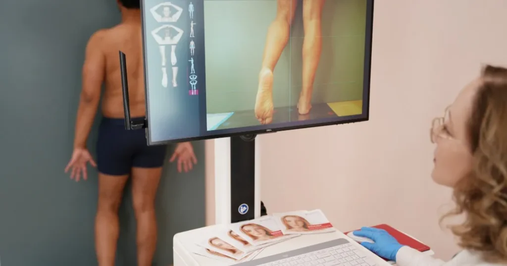
For the skin, synchronized cameras capture thousands of images under controlled lighting. Visible lesions are indexed in a personal skin library so they can be compared during future scans.
Two points are worth understanding before booking. The clinician who discusses the overall scan results is not necessarily a dermatologist. However, Neko states that concerning skin findings are reviewed by a Neko dermatologist and that additional assessment, specialist involvement or referrals may be arranged when needed.
Neko’s U.S. medical terms also explain that it is not a full-service medical practice. Patients are encouraged to continue seeing their treating clinicians for medical diagnoses and conditions identified through the service.
Does Neko Map Moles?
Yes. Neko creates a standardized photographic record of the skin, indexes visible moles and tracks changes over time. This overlaps with total body photography, but it is not identical to a dedicated dermatologist-led mole surveillance visit.
Neko states that spots requiring closer evaluation can be reviewed by a dermatologist within its clinical network. Depending on the finding, additional assessment may be arranged through Neko or the patient may be referred to another provider.
The important distinction is that a broad preventive scan does not necessarily include the same integrated process as a dedicated mole mapping visit:
- A full skin examination performed directly by the dermatologist;
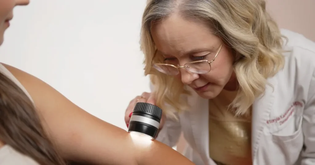
- Lesion-level examination with a handheld dermatoscope;
- Sequential storage of magnified dermoscopic images;
- An immediate clinical decision about whether a lesion should be monitored or biopsied;
- Continuity with the same physician during future surveillance visits.
For someone at average risk with relatively few ordinary moles, a broad preventive scan that includes a skin baseline may be useful. For someone with a personal history of melanoma, numerous atypical moles or a strong family history, a dedicated dermatology pathway provides a more focused level of surveillance.
A useful question to ask any provider is: if one of my moles looks atypical, who examines it with dermoscopy, can the dermoscopic image be stored for comparison, and where would a biopsy be performed if one is needed?
Neko Health Skin Scan vs. Dermatologist-Led Mole Mapping
| Feature | Neko Health Skin Scan | Dermatologist-Led Mole Mapping / TBP |
|---|---|---|
| Primary purpose | Broad preventive-health assessment | Dedicated melanoma and skin cancer surveillance |
| Full-body photography | Yes | Yes |
| Mole indexing | Yes | Yes |
| Initial results review | Overall results reviewed with a Neko clinician | Skin findings reviewed directly by a board-certified dermatologist |
| Dermatologist involvement | Neko states that concerning spots are reviewed by a dermatologist and further assessment may be arranged | The dermatologist performs the examination and interprets the images during the visit |
| Dermoscopy of individual moles | Not presented as comprehensive sequential digital dermoscopy of selected lesions during the base scan | Lesion-level digital dermoscopy performed and stored for future comparison |
| Clinical assessment of a suspicious mole | May require dermatologist follow-up or referral depending on the finding | Clinical and dermoscopic assessment during the same visit |
| Biopsy | Not advertised as part of the standard one-hour scan; separate follow-up or referral may be required | Can be performed during the same visit when medically indicated |
| Longitudinal surveillance | Images stored in a personal skin library for comparison at future scans | Total-body images and selected dermoscopic images compared by the treating dermatologist |
| Areas cameras may not fully capture | May require additional clinical inspection | Scalp, interdigital areas and other difficult-to-photograph sites examined directly by the physician |
| Clinical evidence | Growing service data and observational company reports | Decades of peer-reviewed research, particularly in higher-risk patients |
| Best suited to | Broad health screening with skin imaging as one component | Numerous moles, atypical moles, previous melanoma or significant family history |
Can Neko Health Detect Melanoma?
A scan can identify or flag a lesion that deserves closer examination. It cannot, by itself, establish that the lesion is melanoma.
A flagged spot may ultimately be a benign mole, an atypical but noncancerous mole, another type of skin growth or a skin cancer. Photography and computerized analysis can help determine which lesions deserve attention, but they do not replace clinical examination, dermoscopy or pathology.
When melanoma is genuinely suspected, the definitive diagnosis is generally made by removing an appropriate tissue sample and examining it under the microscope.
What Neko’s Own Data Show
Neko has published real-world findings from tens of thousands of scans performed in Sweden and the United Kingdom. Its 2026 Year Three report includes skin cancers among the diagnoses made following additional assessment and referral. This suggests that the system can surface findings that warrant further investigation.
Neko also describes important limitations of these data. The results come from people who voluntarily chose to undergo scanning, follow-up information was incomplete for some referrals, and the analysis did not include a randomized control group. These are real-world operational findings rather than a formal study establishing the sensitivity and specificity of the skin-imaging system compared with dermatologist-led examination.
What the Dermatology Literature Shows
Total body photography and sequential digital dermoscopy have been studied for decades, particularly in patients at elevated melanoma risk.
A systematic review and meta-analysis involving 41,703 patients evaluated total body photography for melanoma diagnosis. The authors found an acceptable number needed to biopsy among high-risk patients, while also noting substantial variation among the available studies and the need for additional prospective research.
The practical value of longitudinal imaging is greatest in patients with numerous moles or atypical lesions that need to be compared accurately over time. Photography helps identify new or changing lesions, while dermoscopy allows the physician to examine structures within an individual mole that cannot be assessed through an ordinary clinical photograph alone.
Mole Mapping at Dermatology Circle
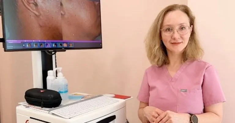
At Dermatology Circle, every step from imaging to clinical decision-making takes place during one private visit with Dr. Viktoryia Kazlouskaya, a double board-certified dermatologist and dermatopathologist.
That second board certification matters. A dermatopathologist is a physician trained to diagnose skin disease by examining tissue under the microscope. Very few dermatologists hold both certifications. This background allows the same physician who examines a mole clinically to evaluate it with an understanding of the microscopic features that distinguish benign, atypical and malignant lesions.
A mole mapping visit at Dermatology Circle combines:
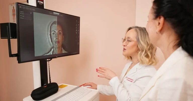
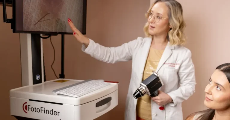
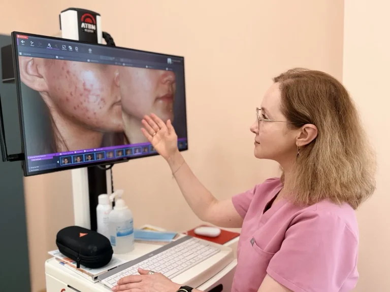
- FotoFinder ATBM total-body imaging, a dermatology-specific system designed for melanoma surveillance and standardized full-body photography;
- Automated change detection and AI-assisted analysis to help identify new or changing lesions between visits;
- Digital dermoscopy of selected individual moles, with magnified images stored for future comparison;
- A full skin examination performed by the physician, including areas that cameras may not adequately capture, such as the scalp and spaces between the toes;
- Same-visit clinical decisions, including reassurance, short-term monitoring or biopsy when medically indicated.
Your images become a longitudinal record that grows more valuable with each visit. The record is interpreted by the same physician who knows your skin, medical history and individual melanoma risk.
Mole mapping is also included in the Dermatology Circle Annual Review, a comprehensive yearly evaluation that can combine total-body skin examination and mole mapping with hair, scalp and cosmetic assessment.
Which One Do You Need?
The two services begin with different questions, so the right choice depends on your goals and individual risk.
A Neko Health Scan May Suit You If:
- You want a broad annual assessment of your heart, blood, circulation and metabolic health;
- You have relatively few moles and no known personal or strong family history of melanoma;
- You want skin photography as one component of a larger preventive-health assessment;
- You are comfortable obtaining additional dermatology care if a lesion requires closer evaluation.
Dermatologist-Led Mole Mapping May Be the Better Choice If:
- You have numerous moles or several that look unusual;
- You have previously been diagnosed with atypical or dysplastic moles;
- You or a close relative have had melanoma;
- You have had other skin cancers or substantial cumulative sun damage;
- A mole has changed, itched, bled or developed another concerning feature;
- You need lesion-level digital dermoscopy and longitudinal comparison;
- You want examination, diagnosis and a management plan coordinated by the same dermatologist.
Some patients may reasonably use both services: a broad preventive-health scan for cardiovascular, metabolic and other health information, together with dedicated dermatologist-led surveillance for the skin.
The choice should follow your medical risk and clinical needs rather than the novelty of the technology.
If melanoma surveillance is your priority, schedule a dermatologist-led mole mapping visit at Dermatology Circle on the Upper East Side.