Two skin growths can both feel rough and look brown or crusted, yet have very different biological significance.
One may be a seborrheic keratosis, a common benign epidermal growth that often requires no treatment.
Another may be an actinic keratosis—a lesion composed of UV-damaged keratinocytes and considered precancerous because some lesions can progress along the pathway toward cutaneous squamous cell carcinoma.
In classic cases, the distinction may be straightforward during a dermatology examination. The difficulty begins when a lesion is pigmented, inflamed, unusually thick, crusted, bleeding or simply does not look like a textbook example.
That is why the question of actinic keratosis vs. seborrheic keratosis cannot always be settled by looking in a mirror or comparing the spot with photographs online.
What Is Actinic Keratosis?
An actinic keratosis, also called a solar keratosis, develops in skin that has accumulated ultraviolet damage.
A keratinocyte is one of the major cell types in the outer layer of the skin. In an actinic keratosis, some of these cells have developed abnormal changes related primarily to chronic UV exposure.
Common locations include:
- Face and forehead;
- Ears;
- Bald or thinning scalp;
- Backs of the hands;
- Forearms;
- Neck and upper chest;
- Other chronically sun-exposed areas.
The clinically important distinction is that actinic keratosis is considered precancerous. Precancerous does not mean that the lesion is already an invasive skin cancer. It means that abnormal cells are present and the lesion has the potential to progress further.
The American Academy of Dermatology explains that actinic keratoses can persist, regress or, in some cases, develop into squamous cell carcinoma.
That does not mean every actinic keratosis will become cancer. It also remains difficult to predict precisely what an individual ordinary-looking AK will do over its lifetime.
What Does Actinic Keratosis Usually Feel Like?
Texture can be one of the most useful clues. Patients often describe an actinic keratosis as:
- Gritty;
- Rough;
- Dry;
- Scaly;
- Sandpaper-like.
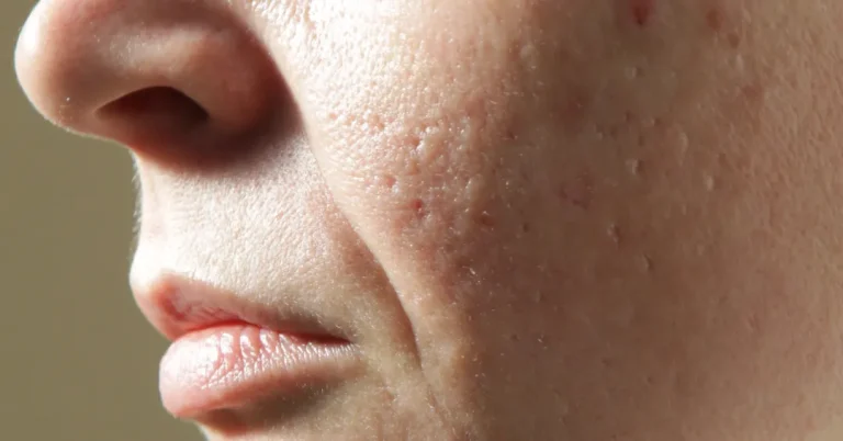
Early actinic keratoses can sometimes be easier to feel than see. Running a finger across the area may reveal a persistent rough patch that is much less obvious visually.
The color is more variable than many patients expect. An actinic keratosis may be skin-colored, pink, red, tan, brown, gray, white or yellowish.
What Is Seborrheic Keratosis?
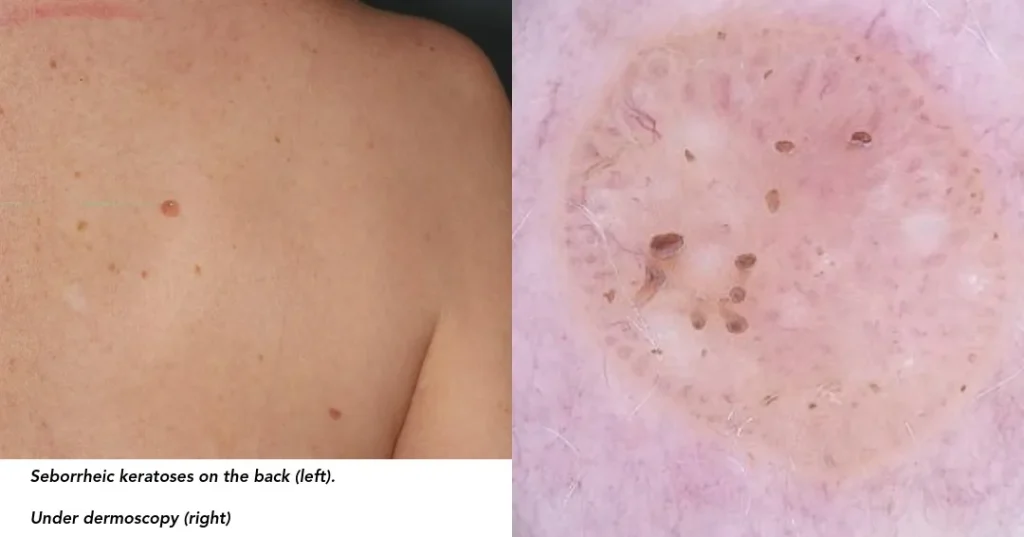
A seborrheic keratosis, or SK, is a common benign epidermal growth. These lesions become increasingly common with age, although they can also appear in younger adults.
Typical seborrheic keratoses may appear:
- Waxy;
- Rough;
- Verrucous or warty;
- Sharply bordered;
- Raised;
- As though they were pasted or “stuck on” the skin.
Their color varies from very light tan to dark brown or black. Some are thin and relatively smooth, while others become thick and markedly textured.
A typical seborrheic keratosis is not a precancerous lesion. Most do not require treatment. The American Academy of Dermatology’s overview of seborrheic keratoses also emphasizes that these common growths are benign despite sometimes resembling precancerous lesions or skin cancer.
Actinic Keratosis vs. Seborrheic Keratosis: Main Differences
| Feature | Actinic Keratosis | Seborrheic Keratosis |
|---|---|---|
| Biological significance | Precancerous keratinocyte lesion | Benign epidermal growth |
| Major association | Chronic UV damage | Age and genetic tendency; not primarily a UV-induced precancer |
| Typical texture | Often gritty, rough or sandpaper-like | Often waxy, rough, warty or “stuck on” |
| Typical locations | Primarily chronically sun-exposed skin | Can occur on many areas of the body |
| Color | Skin-colored, pink, red, tan, brown, gray, white or yellowish | Light tan through dark brown or black |
| Can it be scaly? | Yes | Yes |
| Can it be pigmented? | Yes | Yes |
| Is medical treatment commonly needed? | Frequently | Usually not |
| Can it progress toward SCC? | A subset can progress | Not part of the typical precursor pathway |
| Can it resemble skin cancer? | Yes | Yes |
| Is dermoscopy useful? | Yes | Yes |
| Can biopsy be necessary? | Yes, when clinically indicated | Yes, particularly when the diagnosis is uncertain |
This comparison is useful for understanding common patterns, but it cannot diagnose an individual lesion.
Can a Brown Spot Be an Actinic Keratosis?
Yes. Color alone cannot reliably distinguish an actinic keratosis from a seborrheic keratosis.
This is particularly relevant in more deeply pigmented skin, where relying on the stereotypical image of a bright pink actinic keratosis may be misleading.
The dermatologist evaluates color together with:
- Texture;
- Location;
- Background sun damage;
- Overall morphology;
- How the lesion has changed;
- Dermoscopic findings.
Does Location Help Distinguish AK From SK?
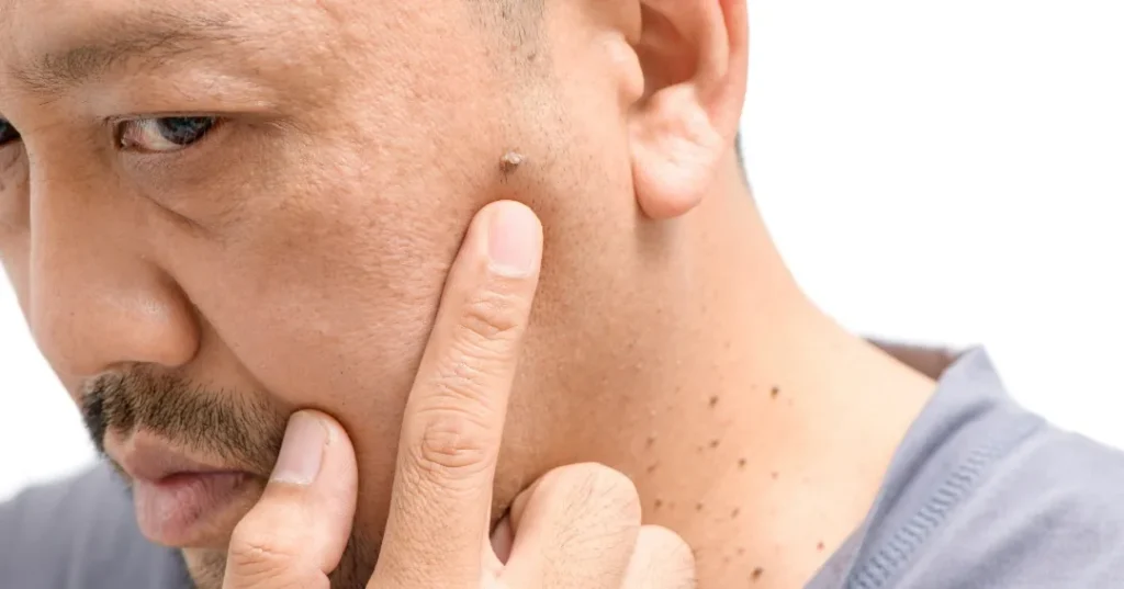
Location provides useful context, but it is not enough to establish the diagnosis by itself.
Actinic keratosis strongly favors chronically sun-exposed areas, especially the face, ears, scalp, forearms and backs of the hands. Seborrheic keratoses can develop on many areas, including the face, scalp, neck, chest and back.
However, both types of lesions can appear on sun-exposed skin. A brown, rough growth on the face cannot be assumed to be an actinic keratosis—or dismissed as a harmless seborrheic keratosis—based on location alone.
Does Every Actinic Keratosis Become Squamous Cell Carcinoma?
No. This point deserves emphasis because the word “precancerous” can unnecessarily frighten patients.
Many actinic keratoses never progress. Some regress spontaneously, some disappear and later recur, and others remain relatively stable for years.
A subset progresses further along the spectrum of keratinocyte dysplasia toward cutaneous squamous cell carcinoma. Current literature continues to describe considerable uncertainty around predicting the behavior of an individual lesion.
An actinic keratosis contains abnormal sun-damaged cells and has enough association with squamous cell carcinoma that dermatologists generally take it seriously and often recommend treatment.
Can Seborrheic Keratosis Turn Into Skin Cancer?
A typical seborrheic keratosis is benign and is not regarded as a routine precursor to skin cancer.
However, a lesion assumed to be an SK may occasionally prove to be something else. In rare circumstances, two different lesions may also coexist in the same location.
The practical concern is usually not that an ordinary seborrheic keratosis transformed into cancer. It is that a skin cancer or atypical lesion can sometimes resemble a seborrheic keratosis clinically.
Can You Reliably Tell AK From SK at Home?
Not always. A classic waxy, sharply bordered seborrheic keratosis may be recognizable, just as a subtle gritty patch on a sun-damaged scalp may strongly suggest actinic keratosis. But many lesions do not follow the classic pattern.
Features that deserve dermatologic evaluation include:
- A new or rapidly changing lesion;
- Repeated bleeding;
- Ulceration or persistent crusting;
- Increasing thickness;
- Tenderness or pain;
- Irregular pigmentation;
- A lesion that looks different from the patient’s other growths;
- A spot that repeatedly returns after being removed or treated.
If the concern is that a lesion is new, evolving, changing color or repeatedly bleeding, our guide to warning signs in a changing skin lesion explains these features in more detail.
How Dermoscopy Helps
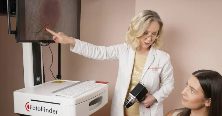
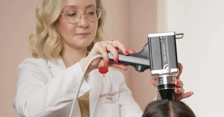
Dermoscopy is a noninvasive examination that uses magnification and specialized illumination to reveal structures that cannot be seen during an ordinary naked-eye examination.
It is particularly useful when two lesions share superficial visual features. Dermoscopy may reveal patterns that support actinic keratosis, seborrheic keratosis, another benign lesion or a possible skin cancer.
At Dermatology Circle, dermoscopy is incorporated when closer evaluation of an individual growth is useful as part of a medical dermatology examination.
Does Dermoscopy Always Provide the Answer?
No. Dermoscopy adds diagnostic information, but it is not infallible.
A thick crust can obscure structures underneath. An inflamed or traumatized seborrheic keratosis can lose some of its typical features. Pigmented actinic keratosis may overlap visually with other pigmented lesions.
If the clinical and dermoscopic findings remain uncertain—or if the lesion has concerning features—a biopsy may be the more appropriate next step.
When Is a Biopsy Necessary?
Most classic actinic keratoses and seborrheic keratoses do not automatically require biopsy.
Biopsy becomes more relevant when:
- The diagnosis remains uncertain after clinical and dermoscopic examination;
- The lesion is unusually thick or tender;
- There is persistent bleeding or ulceration;
- The lesion is rapidly changing;
- Pigmentation is irregular or atypical;
- Squamous cell carcinoma, melanoma or another tumor is part of the differential diagnosis;
- A presumed AK does not respond as expected to treatment;
- The selected removal method should preserve tissue for microscopic evaluation.
Why Histopathology Matters
A biopsy allows part or all of a lesion to be examined microscopically. Histopathology is the microscopic evaluation of that tissue.
When clinical appearance and dermoscopy do not provide enough certainty, histopathology can resolve the question at the tissue level.
This clinical-pathologic correlation is one reason the Dermatology Circle model combines dermatology and dermatopathology within the same physician-led diagnostic approach.
Dr. Viktoryia Kazlouskaya is board-certified in both dermatology and dermatopathology. This background allows the visible characteristics of a lesion to be considered together with its microscopic findings when biopsy interpretation is needed.
What Is Field Cancerization?
A patient with actinic keratosis may have one obvious lesion. Another patient may have numerous rough spots across a chronically sun-damaged scalp, forehead, face, forearms or upper chest.
In that setting, the visible actinic keratoses may represent only part of the UV-damaged tissue. The broader area containing visible and subclinical abnormal keratinocytes is often described as a field of cancerization.
This creates two different treatment concepts.
Lesion-Directed Treatment
Lesion-directed treatment addresses one or several visible actinic keratoses individually.
Field-Directed Treatment
Field-directed treatment addresses a broader area of chronically sun-damaged skin where visible and less obvious abnormal lesions may coexist.
The choice depends on the number of lesions, their location, degree of sun damage, medical history, previous treatments and ability to complete the expected recovery.
How Are Actinic Keratoses Treated?
Actinic keratosis treatment may be lesion-directed, field-directed or a combination of both.
Lesion-Directed Approaches
Selected isolated lesions may be treated with methods such as:
- Cryosurgery using liquid nitrogen;
- Curettage;
- Shave removal;
- Another dermatologist-directed destructive technique;
- Biopsy or complete removal when the diagnosis is uncertain.
Field-Directed Approaches
When a larger sun-damaged area contains multiple lesions, dermatologist-selected options may include:
- Topical 5-fluorouracil;
- Imiquimod;
- Diclofenac;
- Tirbanibulin;
- Photodynamic therapy;
- Selected procedural approaches in appropriate patients.
Different field therapies create different patterns of inflammation, recovery and clearance. The most intensive reaction is not automatically the best treatment for every patient.
Procedures such as selected chemical peels or resurfacing treatments may sometimes be incorporated when broader photodamage and skin texture are also treatment priorities. However, they should not automatically replace established lesion- or field-directed medical therapy for actinic keratoses.
Why Sun Protection Is Part of AK Management
Actinic keratosis is strongly associated with cumulative UV injury. Treating visible lesions while continuing intense, unprotected UV exposure does not address the environmental driver that contributed to their development.
Photoprotection may include:
- Broad-spectrum sunscreen with SPF 30 or higher;
- Regular sunscreen reapplication;
- Wide-brimmed hats;
- Protective clothing;
- Avoiding intentional tanning;
- Reducing unnecessary intense UV exposure;
- Monitoring chronically exposed areas for new or changing lesions.
Sun protection cannot reverse all previous damage, but it can help reduce additional cumulative injury.
When Does Seborrheic Keratosis Need Treatment?
Most seborrheic keratoses require no medical treatment.
Reasons a patient and dermatologist may choose removal include:
- Persistent irritation or itching;
- Repeated rubbing from clothing;
- Catching on jewelry;
- Bleeding after recurrent trauma;
- Cosmetic preference;
- Diagnostic uncertainty.
Seborrheic keratosis is usually treated because it is symptomatic, cosmetically bothersome or diagnostically uncertain—not because it is expected to become cancer.
How Are Seborrheic Keratoses Removed?
Depending on the lesion’s size, thickness, location and pigmentation, dermatologist-directed removal methods may include:
- Cryotherapy;
- Curettage;
- Shave removal;
- Electrosurgical techniques;
- Selected laser treatment.
Each method has trade-offs involving healing time, pigment change, scarring and the possibility of recurrence or incomplete removal.
When the diagnosis is uncertain, the removal method should preserve the opportunity for appropriate histopathologic assessment rather than simply destroying the lesion without obtaining tissue.
What Usually Happens Next?
| Clinical Situation | Possible Next Step |
|---|---|
| Classic benign-appearing SK without symptoms | Often no treatment is necessary |
| Symptomatic or irritated SK | Removal may be considered |
| Typical isolated AK | Dermatologist-directed lesion treatment |
| Multiple AKs in a sun-damaged area | Field-directed therapy may be considered |
| Atypical rough or pigmented lesion | Clinical examination and dermoscopy |
| Persistent diagnostic uncertainty | Biopsy and histopathology may be appropriate |
| Rapidly changing, bleeding or ulcerated lesion | Prompt dermatologist assessment |
This table is educational and does not replace examination of an individual lesion.
Does Mole Mapping Help Distinguish AK From SK?
Digital mole mapping and total-body photography are particularly useful for:
- Documenting numerous pigmented lesions;
- Detecting new lesions;
- Monitoring changes over time;
- Supporting melanoma surveillance in selected higher-risk patients.
Mole mapping is not primarily an actinic keratosis treatment or a substitute for examining a rough lesion with dermoscopy. However, an AK, SK or another lesion may be documented incidentally during total-body photography.
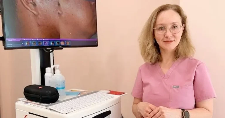
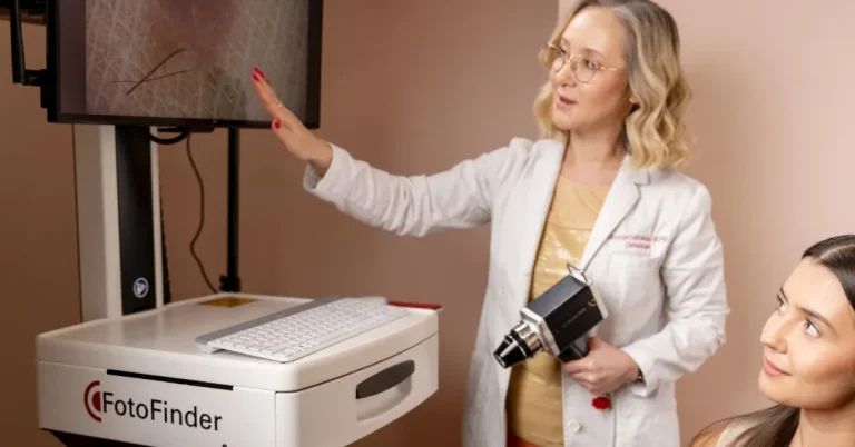
Patients with numerous moles, atypical lesions, a personal history of melanoma or other melanoma-surveillance needs may benefit from FotoFinder mole mapping at Dermatology Circle for those separate indications.
When Should You Schedule an Earlier Examination?
A rough or pigmented lesion should be assessed sooner when it:
- Grows rapidly;
- Becomes painful or tender;
- Bleeds without an obvious injury;
- Develops a persistent open area;
- Changes color or shape;
- Becomes substantially thicker;
- Looks unlike the patient’s other spots;
- Returns repeatedly after treatment.
These features do not automatically mean skin cancer, but they make self-diagnosis less appropriate.
Actinic Keratosis and Seborrheic Keratosis Evaluation at Dermatology Circle
At Dermatology Circle, evaluation begins by determining what the lesion actually represents before selecting a treatment method.
The visit may include:
- Clinical examination by Dr. Kazlouskaya;
- Dermoscopy when additional lesion-level detail is useful;
- Assessment of surrounding sun damage;
- Biopsy when the diagnosis remains uncertain or concerning features are present;
- Histopathologic interpretation when tissue evaluation is needed;
- A lesion-directed or field-directed treatment plan based on the diagnosis.
Dr. Kazlouskaya is board-certified in both dermatology and dermatopathology. This allows the physician who evaluates the lesion clinically to bring the same diagnostic perspective to its microscopic features, helping connect what was seen on the skin with what is found in the tissue.
Once the diagnosis is established, treatment can be selected according to both medical need and cosmetic priorities. Patients with multiple actinic keratoses may require lesion-directed treatment, field therapy or a staged combination plan. A bothersome seborrheic keratosis may be removed using a method chosen according to its location, thickness, skin tone and the desired cosmetic outcome.
If you have a persistent rough, scaly, pigmented or changing spot, schedule a dermatology consultation at Dermatology Circle on the Upper East Side of Manhattan.