If a mole that has looked the same for years and then becomes darker, larger, more irregular, itchy, or simply different from the way you remember it deserves a closer look.
That does not mean every changing mole is melanoma. Moles and other skin lesions can change for benign reasons, including irritation, friction, inflammation, trauma, or gradual changes that occur over time. But change is also one of the clues dermatologists use when deciding whether a lesion needs closer examination.
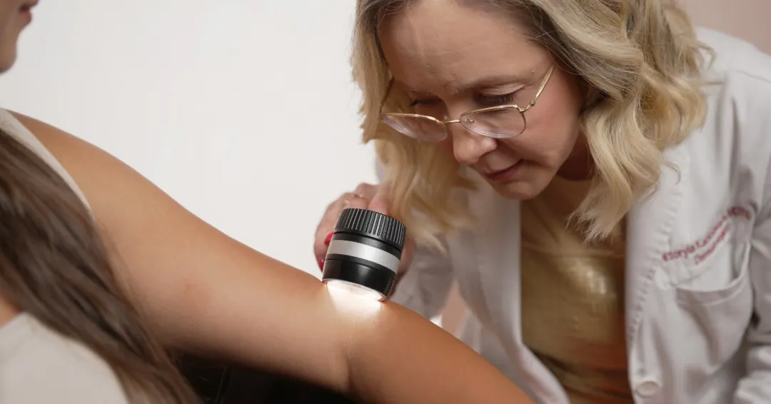
The challenge is that a photograph or checklist rarely tells the whole story. When a dermatologist evaluates a mole, the assessment is not based on one feature alone. It includes how the lesion has changed, how it compares with the patient’s other moles, what it looks like under dermoscopy, and whether the patient’s history changes the level of concern.
For patients who notice a new or changing lesion, a medical dermatology consultation with the skin check is more useful than trying to match it to melanoma photographs online.
Is Every Changing Mole Dangerous?
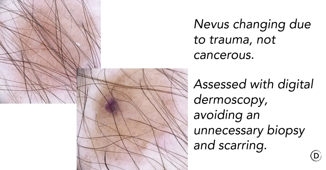
No. A changing mole is not automatically a skin cancer diagnosis. Some benign moles evolve slowly. A lesion may also look different after repeated rubbing from clothing, scratching, sun exposure, or inflammation. Other benign growths can crust, become irritated, or change enough to attract attention.
The opposite assumption, however, can also be risky: deciding that a change is harmless simply because the spot has been present for years.
Melanoma and other skin cancers can reveal themselves through change. What matters is the type of change, its timing, the appearance of the lesion, and how it fits with the rest of your skin.
A practical approach is somewhere between two extremes:
- Do not assume that every changing mole is cancer.
- Do not automatically dismiss a lesion that is clearly evolving.
If you are uncertain, professional examination allows the lesion to be evaluated in context rather than judged from a description alone.
The ABCDEs of Melanoma
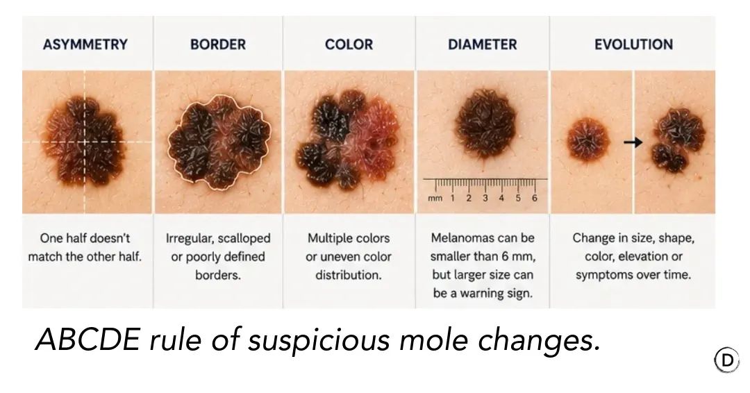
The ABCDE rule is a useful way to remember several common melanoma warning signs. It is a screening tool—not a way to diagnose melanoma at home.
A — Asymmetry
If you mentally divide the lesion in half, one side may look noticeably different from the other.
Some benign moles are not perfectly symmetrical, so asymmetry alone does not establish a diagnosis. It becomes more meaningful when it is new or combined with other unusual features.
B — Border
A concerning lesion may develop borders that are irregular, scalloped, blurred, or difficult to define.
Some benign nevi also have irregular edges. Dermatologists assess the border together with the lesion’s color, structure, history, and dermoscopic pattern.
C — Color
A mole changing color deserves attention, particularly when new shades or uneven pigmentation appear.
Melanoma can contain combinations of brown, black, tan, red, white, or blue, but benign lesions can also have more than one color. The clinical pattern matters more than the presence of a particular color by itself.
D — Diameter
The traditional ABCDE rule draws attention to lesions larger than approximately 6 mm.
Size can be helpful, but it should never be used as a cutoff for reassurance. Melanoma can be smaller than 6 mm. A small lesion that is clearly evolving or looks unlike the patient’s other moles may still warrant evaluation.
E — Evolution
Evolution means change over time. Of all the ABCDE characteristics, this is particularly useful because a lesion may become concerning not because it looked dramatic initially, but because it no longer behaves like a stable mole.
ABCDE provides a framework, but dermatologists also use overall pattern recognition, comparison with neighboring lesions, dermoscopy, and clinical history.
Why “E” for Evolving Deserves Special Attention
There is no single feature that diagnoses melanoma, but change over time is one of the reasons dermatologists pay close attention to a lesion.
Consider a mole that has been stable for many years. If it suddenly becomes noticeably darker, starts expanding at one edge, develops several colors, or begins to look asymmetric, that history gives the examination additional context.
Changes that may justify evaluation include:
- A previously stable mole becoming darker or developing new colors;
- Increasing asymmetry;
- Noticeable enlargement;
- A new raised or thickened area;
- Persistent crusting;
- Unexplained bleeding;
- A change in texture;
- Rapid or continuing growth.
This is also why photographs can be useful. Memory is surprisingly imprecise when trying to decide whether a mole looked the same six or twelve months ago.
A phone photograph can sometimes document a change, but standardized photography is more reliable for longitudinal comparison. In selected patients, digital mole mapping provides a baseline that can be compared with future images.
Photography does not replace examination. Its value is in showing what changed and when.
When ABCDE Doesn’t Apply: Nodular Melanoma
The ABCDE rule was designed around the most common pattern of melanoma, but not every melanoma follows it. Nodular melanoma is a notable exception, and it’s one of the reasons dermatologists don’t rely on ABCDE alone.
Nodular melanoma often looks deceptively unremarkable by ABCDE standards. It can be symmetric, have a single uniform color, and measure well under 6 mm, yet it tends to grow quickly and downward into the skin from an early stage, which makes early detection especially important.
Because this subtype doesn’t fit the standard checklist, some dermatologists use a second mnemonic specifically for it: EFG.
- Elevated — raised above the surrounding skin
- Firm — firm to the touch, rather than soft
- Growing — progressively enlarging over weeks, not years
A firm, raised, growing bump — even one that is a single even color and looks nothing like a “classic” irregular mole — deserves evaluation.
The “Ugly Duckling” Concept
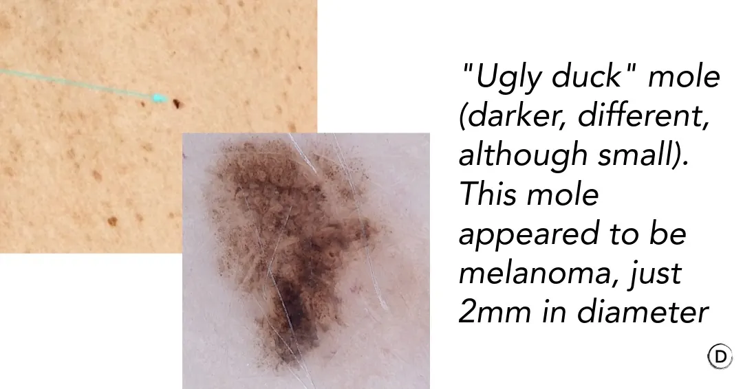
Not every melanoma looks like a textbook ABCDE example. Another clinical strategy is to ask whether a lesion fits the patient’s usual pattern of moles. Most people develop a recognizable personal pattern. Their moles may tend to be small and light brown, or larger and darker, or share similar shapes and dermoscopic features.
Then there may be one lesion that simply does not belong. Dermatologists sometimes call this the “ugly duckling” sign.
Examples include:
- One very dark lesion among many lighter moles;
- One irregular lesion among otherwise round, symmetric moles;
- One pink or lightly pigmented lesion among predominantly dark moles;
- One rapidly changing spot while the surrounding moles remain stable.
An ugly duckling is not automatically melanoma. It is a reason to examine the lesion more carefully. This comparison is particularly helpful in someone with many moles, because evaluating every lesion against an imaginary picture of a “perfect mole” can create unnecessary concern.
Often, the more useful question is: Does this spot look or behave differently from the rest of your skin?
Checking Palms, Soles, and Nails: Commonly Missed Areas
Melanoma can also develop on the palms, soles, and under the nails, in an area of skin that gets little to no UV exposure at all. This subtype is called acral lentiginous melanoma.
Acral lentiginous melanoma is disproportionately more common in patients with darker skin tones, where it represents a much larger share of melanoma diagnoses than in lighter skin. It’s also frequently diagnosed at a later stage, in part because patients and even clinicians don’t always think to examine these areas closely during a routine check.
A few specific things to look for:
- A new or changing dark spot on the sole of the foot or palm of the hand, especially one that doesn’t resolve
- A dark streak or band appearing in a fingernail or toenail
- Hutchinson’s sign: pigment from a nail streak that spreads beyond the nail itself onto the surrounding skin fold, which is considered a significant warning sign for subungual melanoma
- Any nail discoloration, especially in a single nail, that persists or is not explained by trauma
Because these areas are outside of what most people think to check, they deserve a deliberate look during both self-exams and professional skin checks, not just an incidental glance.
What If a Mole Itches or Bleeds?
An Itchy Mole
An itchy mole is not synonymous with melanoma. Itching can result from dry skin, irritation, friction from clothing, eczema, inflammation, or scratching. A benign raised mole may become irritated simply because of where it is located.
What deserves more attention is persistent or unexplained itching in a specific lesion, particularly if the symptom appears together with visible changes in the mole.
A Bleeding Mole
Bleeding also requires context. A raised mole scratched by a fingernail or repeatedly caught on clothing may bleed because it has been traumatized. That is different from a lesion that repeatedly bleeds or crusts without an obvious reason.
If you have a bleeding mole or another lesion that repeatedly develops a scab, arranging an examination is reasonable, especially when there are additional changes in size, shape, color, or surface.
Melanoma is not the only skin cancer dermatologists consider. A lesion that repeatedly bleeds or fails to heal can sometimes represent another type of skin cancer, including basal cell carcinoma. The symptom tells us that the lesion deserves attention. It does not tell us the diagnosis.
What If You Develop a Completely New Spot?
One common misconception is that melanoma always begins inside a mole that has been present for years. It does not.
Melanoma may develop in association with an existing nevus, but it can also appear as a completely new lesion on previously normal-looking skin. This actually happens frequently. That means skin self-examination should not focus only on your existing moles.
Many new spots in adults are benign. Seborrheic keratoses, cherry angiomas, sun-related pigmentation, benign nevi, and other harmless growths appear over time. The goal is not to become suspicious of every new mark. It is to recognize a new and unusual lesion that persists or evolves and have it assessed when appropriate.
Who Benefits From More Frequent Mole Monitoring?
The right frequency of skin examinations is not identical for everyone. More structured surveillance may be considered for people with factors such as:
- Numerous moles;
- Multiple atypical or dysplastic nevi;
- A personal history of melanoma;
- A family history of melanoma;
- Previous atypical biopsies;
- Significant cumulative UV exposure;
- A history of indoor tanning;
- Repeated significant sunburns;
- Immunosuppression.
Someone with relatively few stable moles and no major risk factors may need a very different monitoring strategy from a patient with dozens of atypical nevi and a previous melanoma. This is one reason an individualized plan is preferable to telling every patient that they need the same examination schedule or the same technology.
Not everyone needs digital mole mapping. For the right patient, however, having a consistent photographic baseline can make follow-up much more informative.
Patients with numerous or atypical moles can discuss whether mole mapping in NYC is appropriate for their individual risk profile.
Skin Self-Exams vs. Dermatologist Examination
Self-examination and professional examination serve different purposes.
What Skin Self-Exams Can Do
Looking at your own skin regularly can help you:
- Become familiar with your usual mole pattern;
- Recognize a newly appearing lesion;
- Notice evolution in an existing mole;
- Identify an ugly duckling;
- Bring a specific concern to your dermatologist’s attention.
Do not forget areas that are difficult to see, including the back, scalp, behind the ears, and backs of the legs. A partner or mirror can help with areas outside your direct view.
What a Dermatologist Examination Adds
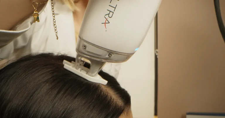
A dermatologist can evaluate features that are difficult to interpret without training. Dermoscopy allows dermatologists to see structures and pigment patterns that are not apparent to the naked eye.
This is why an internet search for “melanoma pictures” is a poor substitute for examination. Benign lesions can look alarming in photographs, while some melanomas can appear surprisingly subtle.
Patients concerned about a particular lesion can start with a comprehensive medical dermatology evaluation.
Skin Check vs. Digital Mole Mapping
Patients sometimes use “skin check” and “mole mapping” interchangeably, but they are not the same thing.
Standard Full Body Skin Examination
A full body skin examination is primarily a clinical evaluation at a particular point in time. The dermatologist examines the skin and evaluates individual lesions, often using a dermatoscope when closer inspection is needed. This can be entirely appropriate for many patients.
Digital Mole Mapping
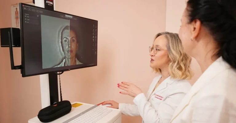
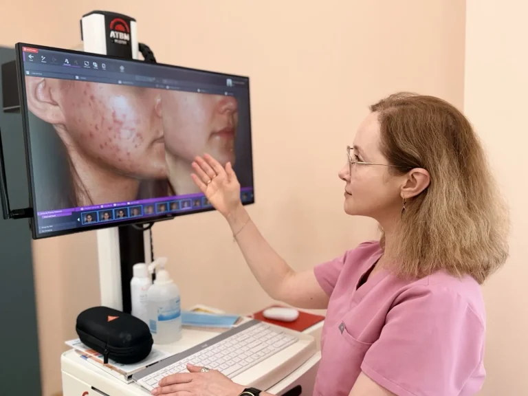
Digital mole mapping adds standardized photography and longitudinal documentation. Instead of relying only on today’s appearance, stored images can be compared with later examinations to help answer two valuable questions:
- Has an existing lesion changed?
- Has a new lesion appeared?
This can be particularly useful in patients with numerous moles, atypical nevi, previous melanoma, or other circumstances in which comparing dozens of lesions from memory would be difficult.
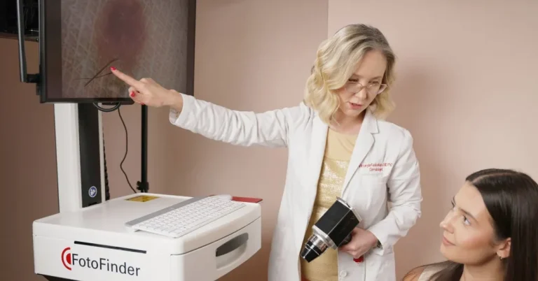
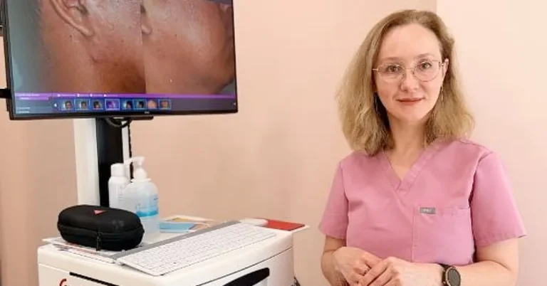
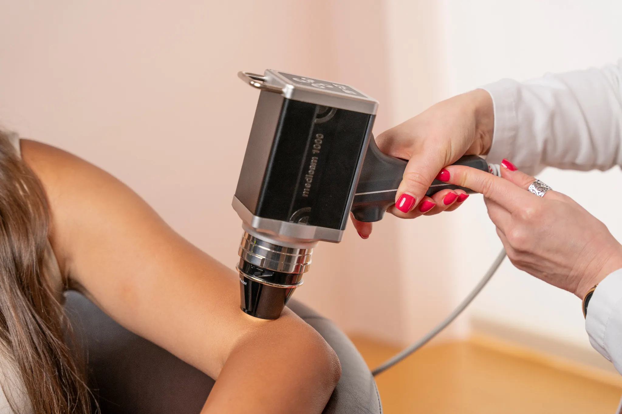
At Dermatology Circle in NYC, mole mapping is performed with the FotoFinder total-body imaging system and combined with dermatologist evaluation. The goal is not to replace clinical judgment with software. The photographs provide another layer of information that can be interpreted alongside the examination.
Not every patient needs this type of surveillance. Mole burden, medical history, previous biopsies, and individual melanoma risk all influence whether monitoring changing moles with digital photography is likely to add meaningful information.
What Happens If a Dermatologist Finds a Suspicious Mole?
Finding a lesion that deserves closer inspection does not automatically mean finding cancer. Several outcomes are possible.
Reassurance. After clinical and dermoscopic examination, the lesion may have features that are consistent with a benign mole or another harmless skin growth. In that situation, no procedure may be necessary.
Monitoring. Sometimes a lesion is not clearly suspicious enough to require immediate removal but is worth following. A dermatologist may recommend photography or another examination after an appropriate interval to see whether the lesion remains stable.
This is one situation in which having high-quality baseline images through digital mole monitoring can be useful.
Biopsy. If the clinical or dermoscopic findings raise enough concern, a biopsy may be recommended.
During a skin biopsy, tissue is removed so that the lesion can be examined microscopically. The exact biopsy technique depends on the type, size, and location of the lesion and the diagnosis being considered.
A biopsy is performed to establish a diagnosis. Being advised to have a biopsy does not mean that a patient already has melanoma. For many lesions, pathology is what resolves uncertainty that cannot be settled by appearance alone.
When Not to Wait for Your Annual Skin Examination
A routine annual visit may be appropriate for some patients, but a clearly evolving lesion does not need to wait simply because your next scheduled skin check is months away.
Consider arranging an earlier dermatologist evaluation if a lesion:
- Changes noticeably in size, shape, or color;
- Develops several new colors;
- Becomes increasingly asymmetric;
- Begins bleeding without clear trauma;
- Develops persistent crusting or a surface change;
- Grows rapidly;
- Becomes an obvious “ugly duckling” compared with your other moles;
- Appears as a new, unusual lesion and continues to evolve;
- Develops a new persistent symptom along with visible changes.
These findings do not mean that you have melanoma. They mean there is enough uncertainty to make examination more useful than continued guessing.
Changing Mole: What the Situation May Mean
| Situation | What It May Mean | Recommended Action |
|---|---|---|
| Stable mole unchanged for years | Often consistent with a benign lesion | Continue routine monitoring appropriate for your risk level |
| Mole changing in size, color, or shape | Change deserves clinical context | Schedule a dermatologist examination |
| New unusual lesion | Many are benign, but a new persistent outlier may require assessment | Do not ignore continued evolution, particularly in adults |
| Bleeding or persistently crusting lesion | Can result from trauma or occur with concerning lesions | Arrange evaluation, especially if unexplained or recurrent |
| Many moles or higher-risk history | Comparing lesions over time can be more difficult | Discuss individualized skin checks or digital mole mapping |
The table is intended as a guide to when to seek evaluation, not as a diagnostic tool.